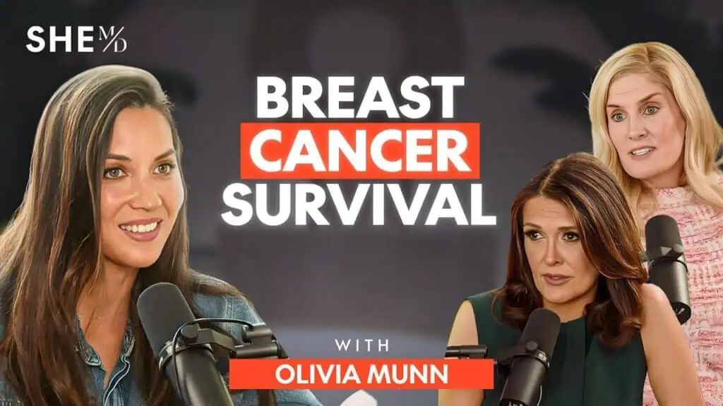
A normal mammogram and negative genetic testing can be reassuring, but they do not answer every question about breast cancer risk. Olivia Munn’s breast cancer diagnosis makes that distinction especially clear. In 2023, after reassuring screening results, a lifetime risk assessment prompted additional imaging that led to the discovery of cancer in both breasts.
The crucial number was approximately 37% lifetime risk of breast cancer. That estimate did not diagnose cancer. It gave Dr. Thais Aliabadi a reason to recommend breast MRI, which set further investigation in motion.
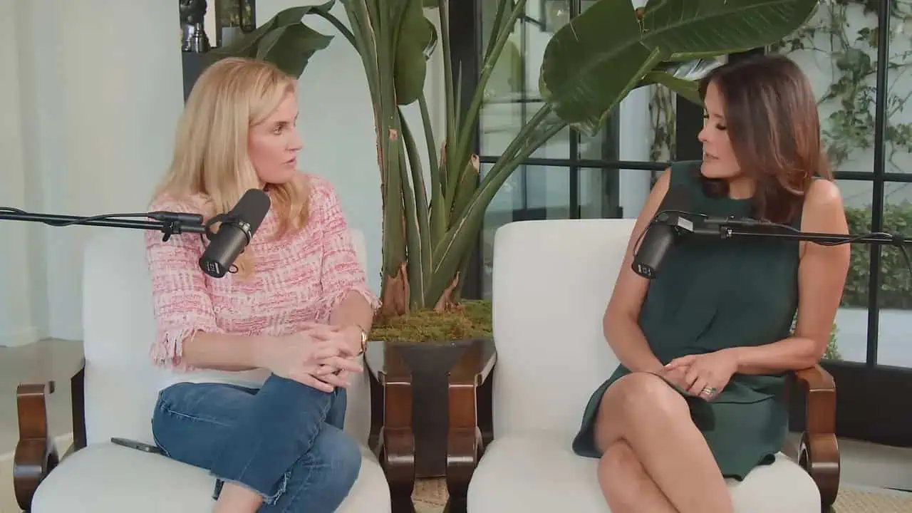
In this interview-style discussion, we explore what Munn’s experience teaches us about personalized screening, dense breasts, genetic testing, double mastectomy, and recovery. The questions and answers below are paraphrased and organized for clarity, with medical explanations distinguished from Munn’s individual experience.
Medical note: We should use this information to prepare for conversations with our healthcare team, not to diagnose ourselves or choose treatment without individualized medical advice.
Table of Contents
- The Breast Cancer Risk Assessment: What It Actually Tells Us
- Normal Mammograms, Dense Breasts, and Additional Imaging
- How Olivia Munn’s Breast Cancer Was Diagnosed
- Double Mastectomy and Reconstruction: Decisions Beyond Detection
- The Emotional Reality of Recovery
- Fertility, Follow-Up, and Self-Advocacy
The Breast Cancer Risk Assessment: What It Actually Tells Us
What was the test that changed Olivia Munn’s breast cancer screening plan?
The turning point was an assessment of her lifetime breast cancer risk. Despite negative genetic testing and a normal mammogram, Dr. Aliabadi calculated a risk of approximately 37%, high enough to recommend additional imaging.
We need to separate two things that are often grouped together: estimating the likelihood of developing cancer and finding cancer that is already present. A risk assessment does the first. Imaging and, when necessary, biopsy help accomplish the second.
In Munn’s case, the estimate changed what happened next. Rather than treating the normal mammogram as the end of the conversation, her doctor recommended breast MRI. An abnormality on MRI led to targeted ultrasound and biopsies.
The practical lesson is not that one questionnaire replaces breast screening. It is that we can use risk information to decide whether our screening plan needs another layer. A number becomes useful when we understand what action it supports.
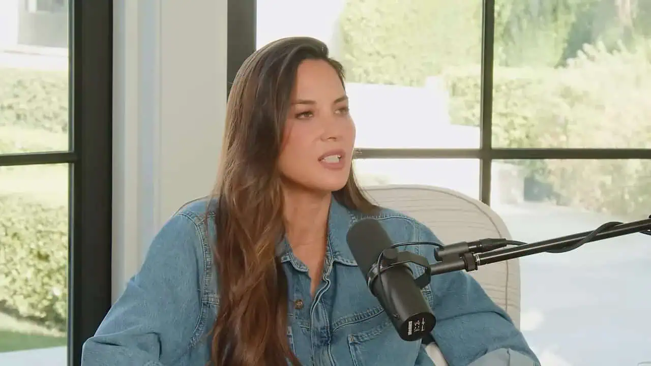
How does a lifetime breast cancer risk calculator work?
We can think of a risk calculator as a structured way to bring several pieces of health history together. The Tyrer-Cuzick model discussed by Dr. Aliabadi considers factors beyond whether a person carries an identified cancer-associated gene mutation.
Depending on the assessment, the information considered can include:
- Age, height, and weight.
- Breast density.
- Age when menstrual periods began.
- Menopausal history.
- Childbearing history, including age at first childbirth.
- Family history of breast cancer.
The result is an estimate, not a prediction of exactly what will happen to us. A high score does not mean cancer is inevitable, and a lower score does not make cancer impossible.
We also need to know what the percentage describes. Some reports estimate risk over a shorter period, while others report lifetime risk. Those figures answer different questions and should not be compared as though they were interchangeable.
For more context, we can review how breast cancer risk assessments work and bring the results to a clinician who can check the information entered and interpret the estimate.
How could Munn have high risk when her genetic testing was negative?
Munn reported negative testing for a broad panel of cancer-associated genes. Dr. Aliabadi also described testing that did not identify a pathogenic mutation. Yet Munn’s overall risk remained elevated.
That is not a contradiction. We should not interpret negative hereditary cancer testing as proof that our breast cancer risk is low. Genetic testing and a clinical risk assessment examine different aspects of risk.
Munn had a family history that included two maternal aunts and a paternal grandmother with breast cancer. Her assessment also considered factors such as breast density and reproductive history. Together, those details mattered even without an identified mutation.
Dr. Aliabadi additionally discussed a risk assessment approach that incorporates small DNA markers alongside clinical information. We should distinguish those markers from a pathogenic mutation in a gene such as BRCA1 or BRCA2.
The question for our clinician is therefore broader than whether a genetic result is negative. We also need to ask what our personal and family history means for screening.
Does everyone need genetic testing before completing a risk assessment?
No. We can begin a clinical risk assessment without first having hereditary cancer testing. A questionnaire and a genetic test are not the same procedure, and they do not necessarily need to happen in a fixed order.
However, family history can make a discussion about genetic testing important. Dr. Aliabadi emphasized looking at cancer history more broadly rather than asking only whether a relative had breast cancer.
She described a patient whose clinical risk estimate was lower than a later estimate that incorporated additional genetic information. That example illustrates why we should ask whether a basic calculator captures enough of our history.
A useful approach is to bring three questions to an appointment:
- Which risk assessment is appropriate for our history?
- Does our family history warrant genetic counseling or testing?
- Would genetic information change our screening recommendations?
We do not need to order every available test ourselves. We need a clear explanation of which information could change our care.
Normal Mammograms, Dense Breasts, and Additional Imaging
Can breast cancer be present after a normal mammogram?
Yes. Munn’s experience shows that a normal mammogram does not rule out every breast cancer. Her earlier mammograms and ultrasounds had been reassuring, but subsequent investigation identified cancer.
Dr. Aliabadi explained that mammography detects many cancers but can miss some, particularly when dense breast tissue makes interpretation more difficult. We should understand that limitation without dismissing mammograms.
Mammography remains an important part of breast cancer screening. The lesson is to ask whether mammography alone is the right plan for our risk level, rather than to abandon it because it is imperfect.
We also should not conclude that Munn’s cancer necessarily developed entirely within the short interval after her normal mammogram. A later diagnosis tells us when cancer was found, not precisely when it began.
Her story gives us a reason to connect screening results with risk assessment. Reassuring imaging and elevated lifetime risk can exist at the same time, and both deserve consideration.
How do we know whether we have dense breasts?
We should check the breast density information in our mammogram report or ask our clinician to explain it. Density is an imaging finding, not something we can reliably determine by touching our breasts.
Dr. Aliabadi described reports using categories such as fatty tissue, scattered fibroglandular tissue, heterogeneously dense tissue, and extremely dense tissue. The wording matters because it can inform a conversation about supplemental screening.
In her practice, she recommends ultrasound in addition to mammography for patients with dense breasts. That is her described clinical approach, not a reason for us to assume every person needs an identical combination of tests.
We can make the discussion more concrete by asking:
- What density category appears in our report?
- How does that finding affect the interpretation of our mammogram?
- How does density contribute to our overall risk estimate?
- Would supplemental imaging be appropriate, and why?
We should leave with an explanation of the plan, not simply the label “dense breasts.”
What is the difference between breast ultrasound and breast MRI?
We should not think of ultrasound and MRI as interchangeable versions of the same test. They provide different information and can serve different roles in screening and diagnosis.
Dr. Aliabadi explained that ultrasound can help evaluate findings such as cysts and clarify some abnormalities identified on mammography. It can also help locate a finding for an ultrasound-guided biopsy.
MRI is a different imaging method that can reveal abnormalities not identified on mammography or ultrasound. In Munn’s case, MRI was the step that raised concern after earlier results had been normal.
Her diagnostic process also demonstrated that visibility can differ between tests. A finding in the left breast could be identified on MRI but could not be located on ultrasound, so an MRI-guided biopsy was needed.
The practical question is not which test is universally best. It is which test helps answer the clinical question we have: screening someone at elevated risk, investigating an abnormality, or guiding tissue sampling.
At what risk level did Dr. Aliabadi recommend breast MRI?
Dr. Aliabadi described 20% or greater lifetime risk as the high-risk category for which she adds breast MRI to the screening plan. Munn’s approximately 37% estimate was well above that threshold.
For estimates between 15% and 20%, she described an intermediate-risk group and a more individualized approach. She also discussed supplemental imaging practices that she considers proactive.
We should distinguish her practice preferences from a universal schedule. The starting age, imaging combination, and frequency need to be discussed with our own clinician rather than copied from another patient’s care.
If our score is elevated, useful questions include whether it changes screening, whether additional testing would refine the estimate, and when the plan should be reviewed.
We can learn more about who needs a breast cancer risk assessment, but an online result should be the beginning of a clinical conversation, not its replacement.
How Olivia Munn’s Breast Cancer Was Diagnosed
What happened after the MRI showed an abnormality?
Munn received a call after the MRI advising that a finding needed further evaluation. Dr. Aliabadi recommended ultrasound, which identified the MRI finding and two additional abnormalities in different areas of the breast.
Those findings led to biopsies. We should be precise about that sequence: the MRI did not provide the final cancer diagnosis. It identified something concerning enough to investigate, and tissue sampling established that it was cancer.
For Munn, the progression from routine screening to diagnostic testing happened quickly. She described having biopsies arranged promptly and then returning to Dr. Aliabadi’s office to receive the results.
We can understand the pathway in four steps:
- Risk assessment: Her elevated estimate supported additional imaging.
- MRI: An abnormality was identified.
- Targeted investigation: Ultrasound found additional areas of concern.
- Biopsy: Tissue testing confirmed cancer.
Keeping those steps separate helps us understand both the value and the limits of each test.

How was cancer found in the other breast?
After cancer was identified in one breast, Munn assembled a specialist team. Surgical oncologist Dr. Armando Giuliano arranged for an experienced radiologist to review the original MRI again.
That second interpretation identified a concerning area in the left breast that had not been mentioned in the first report. Because it could not be located on ultrasound, Munn underwent an MRI-guided biopsy. That biopsy also confirmed cancer.
We should take a measured lesson from this. Imaging interpretation is not infallible, and a specialist review can sometimes clarify findings that matter to treatment planning.
It does not mean every normal study requires another reading. It means that, when a diagnosis raises additional questions, we can ask whether the original images should be reviewed by the treating breast team.
Munn’s eventual diagnosis was bilateral breast cancer, meaning cancer was present in both breasts. The second biopsy resolved an important uncertainty rather than leaving the treatment decision dependent on an assumption.
What type and stage of breast cancer did Olivia Munn have?
Munn described her diagnosis as bilateral luminal B breast cancer. Her doctors characterized her cancer as aggressive and fast-moving, but it was identified at stage 1.
We need to distinguish the cancer’s subtype from its stage. Luminal B describes a type of breast cancer. Stage describes its extent. An aggressive cancer can still be found at an early stage.
Her doctors described the lesions as small, with one close to lymph nodes. We should not translate closeness into a claim that the cancer had spread to those nodes. That is a different question.
The significance of her experience is that additional investigation found an aggressive cancer while it was still at an early stage. We cannot use one person’s account to predict another person’s treatment or outcome.
For broader background, we can consult this breast cancer information resource and ask our care team to explain the exact diagnosis in our own pathology report.
Double Mastectomy and Reconstruction: Decisions Beyond Detection
Why did Munn undergo a double mastectomy?
Munn described several concerns that informed the recommendation: multiple cancerous areas in one breast, findings in different quadrants, an aggressive diagnosis, and subsequent confirmation of cancer in the other breast.
Dr. Aliabadi strongly recommended a double mastectomy and helped connect her with specialists. Munn described receiving different opinions before selecting her team and having the left breast evaluated further.
We should not turn her treatment into a rule for everyone with an elevated risk score. A lifetime risk estimate alone does not mean a person needs breast removal, and Munn’s operation addressed a confirmed cancer diagnosis.
Her surgery took place approximately a month after diagnosis. The urgency was part of her individual treatment plan, not a timeline we should apply to every breast cancer.
When faced with surgical decisions, we can ask what findings support each recommendation, what alternatives are being considered, and which uncertainties need to be resolved before choosing an operation.
What was the nipple-delay procedure she had before mastectomy?
Munn described undergoing a nipple-delay procedure before her double mastectomy. Dr. Aliabadi explained it as a staged surgical approach intended to help preserve blood supply to the nipple area.
Removing breast tissue during mastectomy can disrupt that blood supply. In the approach described, surgeons perform a preparatory procedure about 10 days earlier to allow changes in circulation before the larger operation.
Munn was able to keep her nipples. We should understand that as the result of her surgical plan, not as a guaranteed outcome for everyone.
This gives us another topic to discuss before surgery: whether nipple preservation is appropriate, what the team recommends to support it, and what outcomes remain uncertain.
We should also ask how preservation affects appearance and sensation, since retaining the nipple does not mean the reconstructed breast will feel the same as before.

How did Munn’s tissue expanders differ from her eventual implants?
Munn had tissue expanders after mastectomy and later underwent implant reconstruction. She described the expanders as uncomfortable, firm, high on her chest, and unfamiliar in shape.
We should not judge the final reconstruction by that temporary stage. In her experience, replacing the expanders with implants produced a substantial improvement, although her breasts did not look or feel exactly as they had before.
The conversation contrasted staged reconstruction with direct-to-implant reconstruction. Munn valued having time for postoperative changes to settle before discussing the final implant size.
Dr. Aliabadi, reflecting on her own direct-to-implant experience, said she would have preferred the staged approach because her reconstructed breasts were larger than she wanted. That is a personal reflection, not proof that expanders are always the better choice.
We can use these experiences to ask about tradeoffs: additional procedures, the temporary appearance, the timing of size decisions, and how clearly our preferences can be incorporated into the plan.
Why is reconstruction after mastectomy not the same as cosmetic breast augmentation?
Munn found it difficult when well-meaning friends presented reconstruction as though she were simply receiving a cosmetic breast enhancement. We should recognize why that comparison can minimize what a person has experienced.
A mastectomy removes breast tissue to treat or address cancer risk. Reconstruction happens within that changed anatomy. Munn’s surgeon explained that she should expect changes in appearance and sensation.
Her account included scars, visible implant contours from certain angles, and concern about how clothing would fit. She wanted a smaller result and discussed that preference with her reconstructive surgeon.
We should not generalize every detail of her implant plan to all reconstruction. Instead, we can take away a communication principle: ask what is realistic for our anatomy and describe our priorities clearly.
Survival and appearance are not competing concerns. We can be grateful for treatment while still wanting honest answers about how our body may change.
The Emotional Reality of Recovery
How did Munn handle the shock of diagnosis?
Munn described responding initially by becoming focused on information and decisions. Direct explanations helped her understand what needed to happen next without exaggerated reassurance or unnecessary dramatization.
We should not mistake that focused response for an absence of fear. She described the situation as overwhelming, with medical appointments, treatment decisions, motherhood, and work plans colliding.
A film project was scheduled to begin shortly after she learned she had cancer. Treatment displaced that plan. We can see how a diagnosis affects more than a medical calendar: it can abruptly rearrange family responsibilities and professional commitments.
Her approach was to concentrate on the immediate tasks. That worked for her, but it is not an emotional standard everyone must meet.
We may process a diagnosis with tears, numbness, practical focus, or changing combinations of those responses. The useful goal is to obtain support and understandable information, not to perform strength in a particular way.
What did she find hardest about seeing her body after surgery?
Munn described intense distress when she first saw her breasts with expanders in place. The unfamiliar shape and texture made it difficult to recognize her own body.
We can learn from a mismatch she noticed between the surgical and personal perspectives. Her surgeon was pleased with the technical result, while she was grieving the visible change. Both reactions could exist at once.
She worried about clothing, scars, and how others might interpret the appearance of her reconstructed breasts without knowing her medical history. Those concerns were not simply about vanity. They involved identity, privacy, and feeling comfortable in public.
We should make room for that part of recovery rather than assuming successful surgery resolves every difficulty. Questions about temporary changes, final expectations, and emotional support deserve space in follow-up appointments.
Her later implants looked better to her than the expanders had, but adjustment remained a process rather than a single reassuring moment.

How can we support someone after mastectomy without minimizing the experience?
We can begin by listening rather than trying to supply an immediate positive interpretation. Munn’s account shows that gratitude for being alive can coexist with grief about bodily changes.
Comments that compare reconstruction with elective cosmetic surgery may overlook lost sensation, scars, discomfort, and the emotional meaning of cancer treatment.
More helpful support leaves room for the person’s own perspective. We can ask what feels difficult, what practical help is needed, and whether they want to talk about appearance or would prefer another subject.
We should also avoid imposing ideas about femininity. Munn did not initially understand her breasts as the defining feature of being a woman, but she still felt loss and uncertainty before surgery.
The central principle is to allow complexity. We do not need to persuade someone that a difficult experience has a silver lining before we can be present for them.
Fertility, Follow-Up, and Self-Advocacy
Why did Munn discuss egg freezing as part of this experience?
Munn had frozen eggs at ages 33 and 39, before her breast cancer diagnosis. One influence was hearing about someone who faced an urgent cancer diagnosis and limited time to pursue fertility preservation.
We should treat that story as the motivation behind her decision, not as evidence that every cancer treatment allows the same delay or fertility options.
For Munn, egg freezing created more flexibility around future decisions. Her son Malcolm was conceived without using those frozen eggs, another reminder that a fertility plan and what eventually happens are not always identical.
If future childbearing matters to us, her experience supports raising the subject with our healthcare team rather than assuming it can wait. The options and timing need individualized discussion.
We should not interpret egg freezing as a guarantee of a future pregnancy. Its role in her account was preserving possibilities amid uncertainty.
Does follow-up stop after a double mastectomy?
We should not equate the end of routine breast screening with the end of medical follow-up. The ongoing plan needs to be explained by the treating team.
Dr. Aliabadi described additional imaging she sometimes uses in selected patients, including ultrasound of the axillary lymph nodes and occasional MRI when she is concerned about remaining breast tissue.
She explicitly framed some of those choices as her proactive practice rather than routine indications. We should not copy that schedule without discussing our own situation.
A useful follow-up conversation separates cancer surveillance, postoperative recovery, and concerns related to reconstruction. We can ask who is responsible for each part and what should prompt contact between scheduled appointments.
We should also avoid assuming that a prediagnosis lifetime risk score describes our risk after cancer treatment. That number served a different purpose and requires clinical interpretation in the new context.
What can we do if our concerns are dismissed?
We can start by making the question specific. Instead of requesting every possible test, we can ask how our documented risk score, density, and family history support the proposed screening plan.
We should request copies of relevant reports and ask for unfamiliar terms to be explained. Dr. Aliabadi described a case in which a genetic variant had been confused with a disease-associated mutation. That illustrates the importance of accurate interpretation.
If we still do not receive an adequate explanation, a second opinion can help. Self-advocacy does not require diagnosing ourselves or assuming our clinician is wrong.
We can organize the appointment around three questions:
- What do the findings mean?
- What action do they support?
- What is the follow-up plan if no additional testing is recommended?
The goal is informed, individualized care. We need neither automatic reassurance nor automatic escalation, but a plan whose reasoning we understand.
What practical steps should we take from Munn’s experience?
We can turn awareness into a short preparation checklist:
- Collect our history: Include relevant cancer diagnoses on both sides of the family.
- Find our reports: Keep mammogram, ultrasound, and genetic testing results available.
- Check breast density: Ask what the report says and why it matters.
- Request a risk assessment: Confirm which model is being used and what period the percentage covers.
- Discuss the result: Ask whether it changes imaging or warrants genetic counseling.
- Confirm next steps: Know which appointments or tests need to be arranged.
Dr. Aliabadi also emphasized maintaining a healthy weight, exercising, avoiding alcohol and smoking, and eating a healthy diet. We should understand those habits as part of risk reduction, not a substitute for screening or a basis for blaming anyone who develops cancer.
Munn’s message is ultimately practical: we can complete recommended screening and still benefit from understanding our individual risk. The most useful next step is to connect that risk estimate with a clear, clinician-guided care plan.
Concerned About Your Health? Talk to Dr. Aliabadi
Dr. Aliabadi is an expert OB/GYN who is knowledgeable in all aspects of women’s health and well-being. Dr. Aliabadi and her caring, supportive staff are available to support you through PCOS, endometriosis, menopause, childbirth, infertility, or routine gynecological care. We invite you to establish care with Dr. Aliabadi. Call us at (844) 863-6700 or